1. I would say the three aspects of the assignments for this unit are: My lab report for the Microscope lab. The lab report for the punnet square and dragon lab. I really liked the assignment for Building a Cell.
2. I could have used major improvement on the compendiums. For the first one I was completely lost in thought, and the second one was a little better, although I was still lost in though.
3. I believe my overall grade should be a low A high B. I understand the unit, I was just having problems getting all my ideas out on the compendiums.
4. I'm definitely going to improve on my compendiums. I believe I can develop a better system for it.
Regarding the Unit:
1. I felt really engaged in the unit when we were reading about Cancer and Genetics. I am really interested in Cancer, probably because I'm scared of it. Genetics has always interested me.
2. I was a little distance with work when it came to the Mitosis and Meiosis. I understand it, I just really had a hard time keeping the information.
3. What was helpful to me were the power point slides that Larry Frolich completed for us. I used those tremendously!
4. I was a little confused with the instructions for the Build a Cell project. I was confused with the part for Mitosis, Chromosomes, and the DNA.
5. I was most surprised with the Ethical Issues Essay. When I was reading the articles about cloning, it really opened up my eyes about how cloning could really be a good thing. I'm definitely going to have to research that a little more.
Wednesday, June 11, 2008

Lab Project 1: Building a cell
This project has helped me out in understanding cells and how they go through mitosis, replication, and translation. This lab was completely hands on! Constructing the cell structure was the easiest part of this lab. Trying to figure out how to represent DNA replication, translation, and mitosis was a difficult task. When trying to show mitosis and the translation of DNA helped me understand the process a little easier and gave me more understanding.
While working on this project, here are the items that I used....

Cell plasma: Black licorice
Nucleus: Purple Frosting
Nucleolus: Rice Cake
Nuclear Membrane: Red Licorice
Rough ER: Walnuts
Smooth ER: Blue M&M's
Golgi Aparatus: Twizzlers Pull and Peels
Lysosomes: Yellow Peanut M&M's
Vesicles: Orange Peanut M&M's
Mitchodria: Raspberries
Cilia: Pink Fish Sprinkles
Chromosomes: Black and Red licorice
DNA: Red and Green Sour Candy Strings
Base for DNA: White, Gray, Black, and Blue Fish Sprinkles
Ribosome: Pink Sprinkles
Polypeptides: Turtles

This is the first picture after the cell structure was constructed. This was quite a messy process as my almost 2 year old decided that he likes just about everything in this cell structure :) I used the book model as a reference for all the cell structures.

Here is a picture of the nucleus and chromosomes waiting to get duplicated. The homologous chromosomes come together and line up next to each other, before they come together to form a sister chromatides.

This is a picture representing the sister chromotides. This is part of Mitosis II. They are joined in the center called centromere.
 This was the best way that I could represent the nucleus dividing. As you can see, the chromosomes go to the opposite poles when the nucleous divides.
This picture is representing the DNA structure. As you can see, it resembles a ladder. As you can also see, my camera was having some issues :)

This picture is representing transcription to mRNA.

This is when the mRNA is taken to the ribosome to get translated.
Ethical Essay 1

The article that I read for this assignment is Cloning.
I. Cloning (Saving Endangered animals vs. not have perfection for saving endangered animals)
II. With cloning, this article said that saving endangered animals could be an option. I never really thought about it, but it really could happen. If is possible to create an embryo outside of the womb, and be able to implant into a surrogate mother, than cloning should be able to save endangered animals. The world would never have to worry about having endangered animals which lead to extinction.
III. Although it would be great to save every endangered animal, cloning comes with risks. The cost of cloning is very expensive and it take so much time. There is so much that goes into cloning, and sometimes it doesn't work out. Many of the offspring that are produced from cloning develop a low immunity, which means they can't fight off infection very easily so death comes early in life. Many of the clones that are produced are larger that natural offspring which could lead to some medical problems. Many of them don't live long so they can't report the aging process because they can't get them to live long enough.
IV. Although it would really exciting to see cloning of endangered animals, there is much to be done in order to get it done right. Cloning needs to be just as similar to those that are natural. The knowledge for cloning is there, it just needs a little more perfection. When trying to save endangered animals and they keep dying at early ages, what's the point?
V. In the future I believe that this will be possible. I think that cloning will get perfected in a way that we can help save the endangered animals. I think cloning can be a wonderful thing, although right now it still have a couple errors. If scientists just keep exploring what the difference between the clone and the natural they will be able to come up with a solution and make it possible.
Tuesday, June 10, 2008
Lab 2: Genetics

Here shown is the fly Punnett Square lab. In scenario 5 it asks us to make a cross of
1. a heterozygous,long wing fly
2. a heterozygous, long wing fly
In the Punnett square you can see that the probability of a long winged fly is 75%.
In the Punnett square you can see that the probability of a long winged fly is 75%.

Here is the Dragon lab. In this lab you had to take the first dragon, which happens to be blue, with horns, without scales, with wings, no legs, ect....The second dragon was completely different and you had to change all the genotypes to try to make him identical to the first one.
Genetics are what makes up human beings. When a man and a women produce children, that child is made up of genetics from the parents. Each parent contributes certain features of a child. This could be consisted of (blond/brown/black hair, brown/blue/green eyes, light/dark skin...and so on). You could also look at the fly lab and see that when the two parents decided to have children, what the probability of having a long winged fly would be.
There are a couple terms that were discussed in this lab:
- Genotype: They are the genes of an individual cell. They are represented by two letters. (AA) (Aa) (aa) The Genotypes in the fly lab include (LL)(Ll)(ll). For the dragon lab it included (Hh) (Ss)(Ww) and so on.
- Phenotype: These are what the genes are representing. Examples include (blond hair, blue eyes, dark skin) In the dragon lab (scales, fire, legs, wings, horns, ect..) In the fly lab it included black or grey body, long or short wings.
- Allele: These are the alternative for genes. These are what affect the genes.
- Cross: The Punnett square is an example of this. These are what the parent's give to their children. Cross over.
- Dominant: These are what masks the expression in the allele. They are represented by two uppercase letters (AA) They are heterozygous
- Recessive: These are masked by the dominant genes. They are represented by two lower case letters (aa). They are homozygous
When talking about genetics it may get overwhelming just thinking about it. Using the Punnett Square is an easy way to get a visual representation of what genetics are all about and how they are contributed. It is interesting to try to figure out how genetics work. When I look at my son it's easy for me to picture. If you ever see my son, you'll see that he has some of my features (blond hair, blue eyes, pale skin) and he has some of my husbands features as well (blond hair, pale skin, and height). It is also interesting to see how much he looks like my family members and my in-laws.
I found a great website that has great examples of how to work out the Punnett Square.
http://biology.clc.uc.edu/courses/bio105/geneprob.htm
Lab 1: Microscope Lab
 (This image is a cheek cell at 4x)
When working in biology, especially microbiology, the microscope in an everyday tool. This lab shows us how to use a microscope and how to perfect our skills so we can achieve the goals that we need. We use a microscope to amplify or magnify microscopic organisms to see what they consist of and what their functions are.
There are different parts of a microscope:

- Stage: The stage is the rectangular, flat piece that the slide is placed on. There are clips that are attached to the stage to secure the slide. There are two knobs that are associated with the stage. They move the stage up and down and left to right.
- Focus knobs: There are two focus knobs. The bigger knob, or coarse knob, moves the stage quickly. The smaller knob, fine knob, moves the stage more slowly and is more for smaller adjustments.
- Iris: Is what helps with the transparency of the specimen. It is located under the stage.
- Oculars: These are what we look through. You can move them to adjust to your eyes. If you don't adjust them correctly, you will see double so make sure you only see one image.
- Objectives: These are what magnifies the images. On the simulator we used there were 4 objectives (4x, 10x, 40x, and 100x). To switch to the different objectives you just rotate them to the objective you want to use. Make sure when you are adjusting them, you have enough room between the lens and the stage! Otherwise, you will scratch the lens and the stage!
When you put a specimen on the stage you want to make sure it is centered and over the hole on the stage. The light needs to be able to shine through the specimen so you can get a clear image. Set the objective to the right magnification that you want. Use the focus knobs to adjust the specimen to the right image that you want. If you want to adjust it quickly, use the coarse knob. If you want to make smaller adjustments, use the fine knob. The xy controls are used to move the specimen side to side, or left to right.
Working with a microscope takes a lot of work and practice. I found that the more I used it, I became more familiar with it. I would use different objectives to see what images I would see and how they would differ from each other. Just remember, the more you utilize it, the better you will get with it. Practice makes perfect!
Some historical facts:
Zacharias Janssen was the first to develop a microscope in 1595. Many people believe that his father, Hans, is the one who actually developed the first microscope
In 1660, Robert Hooke took the microscope and modified it. He was a very mathematical person and used his knowledge to improve the microscope.
"Anton van Leevwerhoek was the first person to ever describe bacteria (from teeth scrapings), protozoans (from pond water) and helped prove the theory of blood circulation"

(this cheek cell at 10x)


(cheek cell at 100x)
Sunday, June 8, 2008
Compendium Two: Genetics
For this section, we covered 4 chapters of the book; Patterns of Chromosome Inheritance, DNA Biology and Technology, Cancer, and Genetic Inheritance. We went from the cell cycle to how abnormalities in the cell cycle can cause certain diseases. As a nursing major, I am really into how diseases are caused and what we can do to try and stop them from occurring.
Chapter 18:
--Humans have 46 chromosomes in 23 pairs...22 pairs are autosomes, 1 pair are sex chromosomes
Cell Cycle has two phases..
1. Interphase (90% of cell cycle---about 20 hours)
- G1- doubles in organelles
- S- DNA replication, duplication of chromosomes
- G2- Growth
2. Cell division
- M- Mitosis
Mitosis is when the parent cell (original) produces daughter cell (duplication)
The process of Mitosis
- Prophase (centrosome duplicates and separates)
- Metaphase (chromosomes are at the center)
- Anaphase (Separate and move to opposite ends)
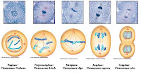
- Telephase (at opposite ends)
( http://www.biology.iupui.edu/biocourses/N100/2k3ch8mitosisnotes.html)

Meiosis is when the daughter cells divide...so you'll have 4 daughter cells total after the division
- Meiosis I- Line up side by side, paired chromosomes at equator,
- Meiosis II- Zygote (1st new individual)
The stages of Meiosis Prophase I
- Metaphase I
Mitosis vs. Meiosis
- Meiosis requires 2 nuclear divisions, Mitosis only requires 1
- 4 daughter cells for meiosis, mitosis only 2
Spermatogenisis and Oogenisis are the production of sex cells (sperm and egg)
Chapter 21:
The structure of DNA is it "resembles a ladder" (Sylvia Mader, Human Biology 10th Edition). The middle part of the "ladder" is contained of 4 different materials. (A pairs with T: G pairs with C)
When the replication of DNA occurs, only one strand is original. It will duplicate itself to get the other strand.

The structure of RNA differs from DNA because it only has one strand instead of the double helix. Instead of A, T, G, and C...RNA is constructed of A, U, C, and G.
- rRNA joins with proteins and is produced in nucleus
- mRNA is also produced in nucleus and is Messenger RNA
- tRNA must have at least 20 present to keep functioning
Transcription is the process of
- Forming mRNA
- Processing mRNA
- Translation
Chapter 19
According to the Cancer webpage (http://www.cancer.org/) there was an estimated 1,444,920 cases of new cancer in the United States alone. Cancer occurs when a cell does not have the capability of dying(apoptosis). It will just keep regenerating.
Cancers have abnormal nuclei they are larger than others and have a different number of chromosomes.
When cancer cells form they just keep growing until they produce a mass, called a tumor. Cancer cells "pile on top of each other" (Sylvia Mader, Human Biology 10th Edition). Benign tumors are usually contained and are tight together. Cancer usually grows all over the place with no real shape.
Three different stages
- Initiation
- Promotion
- Progression
Cancer can often be a genetic disease. p53 is a protein that stops the cell cycle. A lot of cancers are lacking the p53 protein.
There are different types of cancer
- Prognosis (tumor has spread to tissues and other parts of body)
- Carcinomas (Skin, breast, liver, pancreas, intestines, lung, prostate, thyroid)
- Sarcomas (bone, fibrous)
- Leukemias (blood)
- Lymphomas (lymphatic system)
Cancers, aside from genetics, can also be caused by the environment. Radiation, organic chemicals, and diet can have an effect on cancer.
You have a better chance of going into remission if you follow these 7 steps
- Change in bowel or bladder
- A sore that doesn't heal
- Unusual bleeding
- Thickening or lump

- Indigestion or difficulty swallowing
- Obvious change in move/wart
- Nagging cough or hoarseness


(Page 411 from our Text book, Sylvia Mader Human Biology 10th edition)
There are many different ways to handle cancer and hopefully go into remission
- Surgery
- Radiation
- Chemotherapy
- Bone Marrow transplant
Newer technology
- Immunotherapy
- p53 gene therapy
Chapter 20:
Genotype are the genes of the individual. Dominant genes are represented with 2 upper case letters(AA), recessive genes are represented with 2 lower case letters(aa), and heterogenius is represented with 1 upper case and 1 lower case(Aa) (although it takes the characteristics of Dominant genes)
The product that we are testing (ex: blond hair) is called the phenotype
When trying to figure out what the percentage of a phenotype you can use the Punnett Square. Here is an example


When forming a family pedigree you can set it up like this:

When the environment changes some of the outcomes of genetics is it called Polygenic inheritance. (skin color can be changed by the sun)
Example of Incomplete dominance is when someone with curly hair and another person with straight hair produce a child with wavy hair. (Sylvia Mader, Human Biology 10th edition)
Thursday, June 5, 2008
Compendium One
Compendium One: CELLS
This first unit is about cells and how they are incorporated into all life. It also shows that all cells are contained of atoms and how they are bonded to form many different organs and other units. In chapter three, the book shows the different parts to a cell..and you can relate some of the parts of a cell to human organs. The cell has a “powerhouse” or what I refer to is the “brain” and many other parts that can translate into human organs.
Cells are made up of atoms and atoms are made up of:
1. Protons- which have a positive charge and are inside the nucleus
2. Neutrons- have a negative charge and are inside the nucleus
3. Electrons- circle around the nucleus
When you are trying to put together two atoms and 1 atom has one less electron and have an opposite attraction you get ionic bonding.
When you try to put two atoms together that have an even number of electrons and are attracted together you get covalent bonding.
This first unit is about cells and how they are incorporated into all life. It also shows that all cells are contained of atoms and how they are bonded to form many different organs and other units. In chapter three, the book shows the different parts to a cell..and you can relate some of the parts of a cell to human organs. The cell has a “powerhouse” or what I refer to is the “brain” and many other parts that can translate into human organs.
Cells are made up of atoms and atoms are made up of:
1. Protons- which have a positive charge and are inside the nucleus
2. Neutrons- have a negative charge and are inside the nucleus
3. Electrons- circle around the nucleus
When you are trying to put together two atoms and 1 atom has one less electron and have an opposite attraction you get ionic bonding.

When you try to put two atoms together that have an even number of electrons and are attracted together you get covalent bonding.

Organic Molecules
1. Carbohydrates consist of simple carbohydrates, complex carbohydrates, and fiber
2. Lipids are fats to animals, and oils to plants. They contain more energy
3. Proteins have amino acids and have an unusual shape that is necessaryfor continuation.
4. Nucleic Acids consist of DNA and RNA and uses ATP to transport energy.
DNA forms helix RNA does not for helix
1. Carbohydrates consist of simple carbohydrates, complex carbohydrates, and fiber
2. Lipids are fats to animals, and oils to plants. They contain more energy
3. Proteins have amino acids and have an unusual shape that is necessaryfor continuation.
4. Nucleic Acids consist of DNA and RNA and uses ATP to transport energy.
DNA forms helix RNA does not for helix
Cells are very interesting in the fact that new cells are produced from cells that are already present. Some of the structures of the animal cell can almost be related to a human being.

1. Plasma membrane: think of it as human skin because it is the outermost part of the cell. In the plasma membrane there are a couple of processes that take place
“Diffusion is the random movement of molecules from the area of highest concentration to the area of lower concentration, until they are equally distributed” (Sylvia S. Mader, Human Biology 10th Edition)
Osmosis is the diffusion of water across the plasma membrane
Active transport is movement from lower to higher concentrations and requires proteins
2. Nucleus you can think of as a humans heart. It keeps the cell going. The nucleus keeps the cells genetic information.
3. Endoplasmic Reticulum has two different parts the Rough ER is where proteins are processed and modified and the Smooth ER processes and transports to the plasma membrane or the Golgi Apparatus.
4. Golgi Apparatus and lysosomes you can think of as the digestive system. It is where proteins and lipids are processed, packaged and secreted.
5. Mitochondria are like the brain of a cell. It processes ATP for energy and keeps the cell going.
There are four different types of tissues.
1. Connective- connects (tendons, bone, ligaments)
Fibrous, supportive, bone, and fluid tissues
Fibrous, supportive, bone, and fluid tissues
2. Muscular- moves the body (muscles of the body, heart)
Skeletal, smooth, and cardiac
Skeletal, smooth, and cardiac
3. Nervous- conducts impulses (brain)
Neurons and Neuroglia
Neurons and Neuroglia
4. Epithelial- covers the body (skin)
Simple, Columnar, Cuboidal, Squamous, Pseudostratified, Transitional, Stratified.
Simple, Columnar, Cuboidal, Squamous, Pseudostratified, Transitional, Stratified.
Figure 4.12 (page 74-75) goes into all the different systems which has a good representation of how each system works.
Cells are a part of life. They are so tiny, but are so important. They are what make up man, animals, plants....just about everything! Every little thing about cells; how they bond, how they work, and how they make up objects is amazing.
Subscribe to:
Posts (Atom)